
HCC
Primary liver cancer
Hepatocellular carcinoma begins in liver cells and often develops in a liver already affected by cirrhosis or chronic disease. Imaging pattern and liver reserve are central to planning.
Cancer care · Interventional oncology
Selected primary and metastatic liver tumors can be treated through a tiny access point or precisely targeted under imaging. The first question is whether one of those options fits your diagnosis, liver function, anatomy, and broader cancer plan.
Already have a CT or MRI? Bring the images and report. A specialist can review them in the context of your oncology plan.
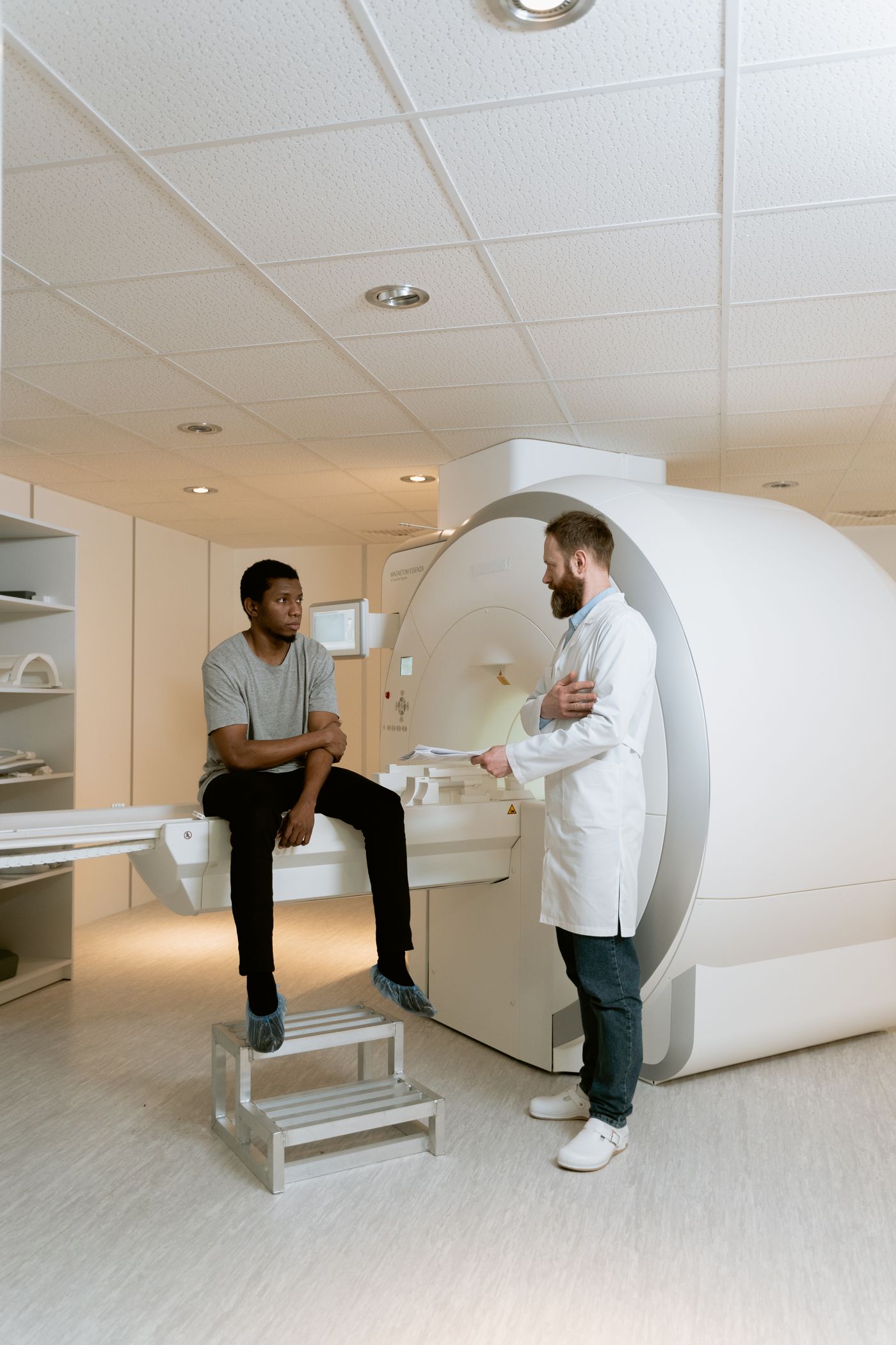
A spot on the liver can represent a primary liver cancer, a cancer that spread from somewhere else, or a noncancerous lesion. Even after cancer is confirmed, the best treatment depends on how much liver is involved, how well the liver is working, whether disease is present elsewhere, and what the full care team is trying to accomplish.
EVSLA evaluates the image-guided part of that decision. A consultation may lead to liver-directed treatment, additional workup, coordination with another specialist, or the conclusion that an interventional procedure is not the right next step.
Clinical context: National Cancer Institute · AASLD · RadiologyInfo
Hepatocellular carcinoma begins in liver cells and often develops in a liver already affected by cirrhosis or chronic disease. Imaging pattern and liver reserve are central to planning.
Cholangiocarcinoma that begins inside the liver has a different biology and treatment pathway. Liver-directed therapy may be considered only in selected situations.
These began elsewhere—such as the colon, breast, lung, or neuroendocrine system. The original cancer type and disease beyond the liver shape the plan.
Early liver cancer may not cause obvious symptoms. Many tumors are found during surveillance, imaging for another concern, or follow-up of a different cancer.
People with cirrhosis or certain chronic liver conditions may have regular ultrasound and blood testing that identifies a new concern.
A liver lesion may appear on ultrasound, CT, MRI, or PET imaging ordered for another symptom or health question.
Upper-right abdominal discomfort, fullness, low appetite, unintended weight loss, fatigue, or jaundice can prompt evaluation—but none is specific to cancer.
Imaging may identify liver lesions during staging, treatment response assessment, or surveillance for a cancer that began elsewhere.
Contact your cancer or liver team promptly for new or worsening jaundice, confusion, fever, vomiting blood, black stools, or severe abdominal pain. Call 911 for severe or life-threatening symptoms.
A procedure can be technically possible and still not be the right treatment. EVSLA reviews the images and clinical context together before recommending a path.
Request a Case ReviewMultiphasic contrast CT or MRI can help characterize a lesion, map its relationship to vessels and bile ducts, and show the number and distribution of tumors. Prior studies help reveal change over time.
Some hepatocellular cancers can be diagnosed from characteristic imaging in at-risk patients. Other lesions may require biopsy or review of pathology from the cancer’s original site.
Laboratory results, cirrhosis or portal hypertension, symptoms, kidney function, medications, and general health help determine what the liver and the patient can safely tolerate.
The goal may be cure in a highly selected case, local control, downstaging, bridging to transplant, symptom relief, or support of a broader systemic treatment plan. The role of interventional radiology changes with that goal.
Y90, microwave ablation, and TACE are not competing versions of the same treatment. Each approaches the tumor differently, and each has its own planning, safety, and follow-up requirements.
A small catheter is guided into the liver’s arterial supply to deliver radiation-containing microspheres close to selected tumors while limiting exposure to the rest of the body.
A primary or metastatic liver tumor is suited to arterial treatment and the mapping study, vascular anatomy, liver reserve, and broader oncology plan support it.
Using CT or ultrasound guidance, a specialist places a thin antenna into the tumor and uses heat to destroy targeted tissue without removing part of the liver.
There are a limited number of smaller tumors in locations that can be safely reached and treated while protecting major vessels, bile ducts, bowel, and diaphragm.
Transarterial chemoembolization delivers chemotherapy into the artery feeding a tumor and then reduces that blood flow, concentrating treatment in the liver.
Selected liver-dominant tumors—commonly certain hepatocellular cancers—are not suited to surgery or ablation and liver function is adequate for embolization.
No single scan measurement decides eligibility. A specialist combines imaging, laboratory results, prior care, and the intended treatment goal.
A focused review is most helpful when the specialist can see the actual images and understand what the rest of your care team has recommended.
Tell the scheduling team your diagnosis, where you are in care, and which records are available.
The specialist evaluates the tumor pattern, anatomy, liver reserve, and treatment context.
When appropriate, EVSLA aligns the interventional plan with your oncology or liver team.
You receive a clear explanation of fit, alternatives, preparation, recovery, and follow-up.
A consultation is a decision-making visit. It does not commit you to a procedure.
EVSLA’s board-certified interventional radiologists evaluate the imaging, explain whether a liver-directed option may fit, and coordinate next steps with the clinicians already treating your cancer.

His specialty training includes oncology and other image-guided treatments, with a direct, steady approach to complex decisions.
Meet Dr. Dermendjian
He brings clear communication and a collaborative approach to complex vascular and minimally invasive image-guided care.
Meet Dr. Kansagra
His UCLA fellowship training and interventional oncology experience support careful planning for selected embolization and ablation cases.
Meet Dr. MathevosianYou do not need to choose a physician before requesting care. EVSLA will help route the case based on the clinical question, records, and availability.
Choose the clinic most convenient for your evaluation. Procedure location and availability depend on the treatment and clinical need, and the scheduling team will confirm the appropriate site.


1407 Foothill Blvd
La Cañada Flintridge, CA 91011
Clear answers about diagnosis, eligibility, coordination, and what to bring to a consultation.
No. Some liver lesions are benign, and some cancers are best observed or treated in another way. The right next step depends on what the lesion is, how much of the liver is involved, liver function, overall cancer stage, symptoms, and goals.
A specialist reviews the cancer type, number, size and location of tumors, liver function, vascular anatomy, disease outside the liver, prior treatments, and the larger oncology plan. These procedures are not interchangeable, and some patients are better served by surgery, transplant, medication, radiation, observation, or supportive care.
Primary liver cancer begins in the liver. Metastatic liver cancer began in another organ and later spread to the liver. That distinction changes staging, treatment goals, and which specialists and therapies should be involved.
Not always. In some at-risk patients, hepatocellular carcinoma can be diagnosed using characteristic findings on multiphasic CT or MRI. Other lesions may require tissue diagnosis. Your team decides based on the imaging, medical history, and how the result would change care.
No. Image-guided liver treatment is usually one part of multidisciplinary care. EVSLA can review the interventional radiology options and coordinate recommendations with oncology, hepatology, surgery, transplant, radiation oncology, and other members of your care team as appropriate.
Bring the actual CT, MRI, or PET images when possible, not only the reports. Also bring pathology results, recent laboratory results, a medication list, prior cancer treatment records, and the questions you want answered.
Some ablation procedures may be used with curative intent for carefully selected small tumors. Other liver-directed treatments may be used to control disease, reduce tumor burden, bridge to transplant, support downstaging, or relieve symptoms. The intended goal and expected benefit must be individualized.
Coverage depends on your plan, network, diagnosis, medical necessity, referrals, and prior authorization requirements. EVSLA can review available benefit information, but verification is not a guarantee of payment.
Share where you are in the process and what imaging you have. EVSLA can help coordinate an evaluation and explain what records are needed before the visit.