
RCC
Suspected or confirmed renal cell carcinoma
The subtype, stage, size, growth, and location help shape a plan that may include surgery, surveillance, ablation, radiation, or medication.
Cancer care · Interventional oncology
Some small, localized kidney tumors may be treated with an image-guided needle probe. The first question is whether ablation fits the diagnosis, tumor location, kidney function, surgical options, and the rest of your care plan.
Already have a CT or MRI? Bring the images and report. A specialist can review the mass in the context of your kidney health and urology or oncology plan.
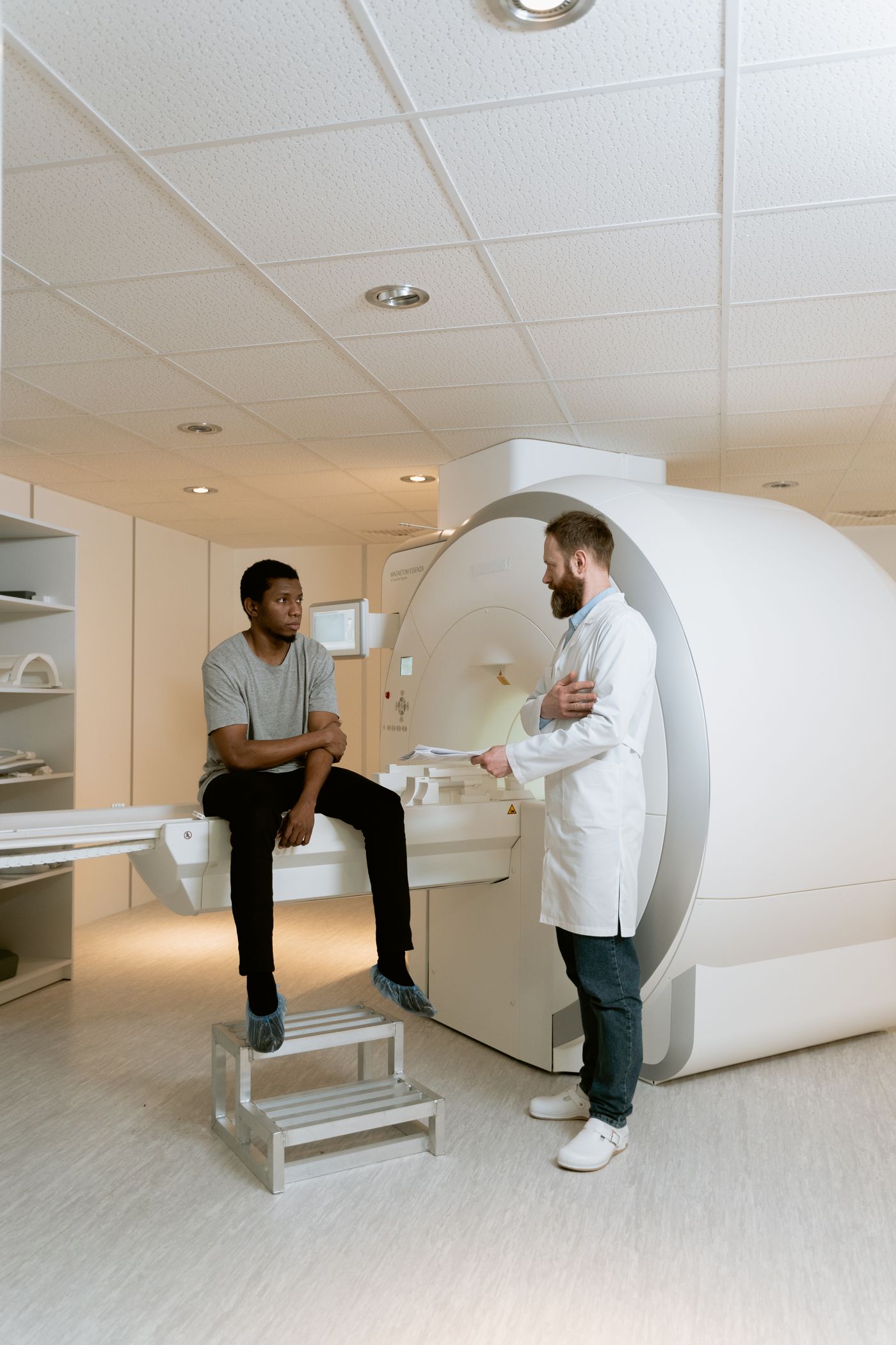
“Kidney mass” describes an imaging finding, not a final diagnosis. A mass may be renal cell carcinoma, a benign growth, or something that cannot be classified from the first scan alone. Tumors that began elsewhere and spread to the kidney are evaluated differently.
For localized kidney cancer, surgery—often removal of the tumor while preserving the rest of the kidney—remains a principal treatment. Active surveillance and image-guided ablation can also be appropriate for selected patients and tumors.
Clinical context: National Cancer Institute · American Urological Association · RadiologyInfo
The subtype, stage, size, growth, and location help shape a plan that may include surgery, surveillance, ablation, radiation, or medication.
Additional imaging, comparison with older scans, biopsy, or surveillance may be more useful than moving directly to treatment.
When cancer has spread, the role of local treatment changes. Medical oncology, urology, radiation oncology, and interventional radiology may all contribute.
A renal mass often appears on imaging ordered for another reason. Symptoms can occur, but symptoms alone cannot determine whether a mass is cancer.
An ultrasound, CT, or MRI for abdominal pain, an injury, or another condition may reveal a mass unexpectedly.
Visible or microscopic blood in the urine can have many causes. It deserves medical evaluation rather than self-diagnosis.
Persistent side or back discomfort, anemia, weight change, or other findings may prompt testing, but they are not specific to kidney cancer.
A known small renal mass, hereditary risk, or prior cancer may lead to scheduled imaging and comparison over time.
Contact your clinician promptly for visible blood in the urine, persistent flank pain, fever, or other concerning changes. Call 911 for severe bleeding, fainting, chest pain, trouble breathing, or another emergency.
A safe recommendation depends on more than tumor size. EVSLA reviews the actual imaging and the clinical context before discussing whether an image-guided procedure belongs in the plan.
Request an EvaluationContrast-enhanced CT or MRI helps define the mass, growth pattern, stage, and relationship to vessels, bowel, the urine-collecting system, and other organs. Prior images add important context.
Imaging can strongly suggest kidney cancer, but it cannot identify every mass with certainty. A renal mass biopsy may help avoid treating a benign lesion and guide the treatment strategy.
Kidney function, whether both kidneys are working, prior kidney surgery, medications, bleeding risk, heart and lung health, and other conditions affect what can be done safely.
The discussion should include partial or radical nephrectomy, active surveillance, ablation, radiation, medication, or a combination as appropriate—not just the procedures an interventional radiologist can perform.
Kidney tumor ablation uses imaging to guide one or more thin probes into the mass. Cryoablation freezes the target; microwave ablation heats it. Neither is automatically the better choice.
Using CT or ultrasound guidance, a specialist places thin probes into the tumor and creates a controlled ice zone intended to destroy the targeted tissue.
A smaller, localized tumor can be safely reached and the visible ice zone can be monitored while protecting nearby bowel, ureter, vessels, and healthy kidney tissue.
A thin antenna is placed into the mass under imaging guidance and uses microwave energy to create a controlled zone of heat within the targeted tissue.
Tumor size, position, access, and nearby structures support heat-based treatment and the clinical team believes it offers an appropriate balance of control and risk.
A technically reachable tumor is not automatically an ablation case. The expected cancer outcome, long-term evidence, kidney reserve, age, health, and personal priorities all matter.
Partial or radical nephrectomy, active surveillance, stereotactic radiation, systemic treatment, or another multidisciplinary plan based on diagnosis and stage.
No single scan measurement determines eligibility. A specialist combines the imaging, kidney reserve, biopsy information, alternatives, and treatment goal.
A focused review works best when the specialist can see the actual images and understand what your urologist, oncologist, or primary clinician has already recommended.
Tell the scheduling team how the mass was found, where you are in care, and which records are available.
The specialist evaluates the mass, access route, nearby structures, kidney reserve, and stage.
Ablation is weighed against surgery, surveillance, and other relevant care—not considered in isolation.
You receive a clear explanation of fit, alternatives, preparation, recovery, and follow-up.
A consultation is a decision-making visit. It does not commit you to a procedure.
EVSLA’s board-certified interventional radiologists review the imaging, explain whether ablation may fit, and coordinate next steps with the clinicians already managing your kidney or cancer care.

His specialty training includes oncology and other image-guided treatments, with a direct, steady approach to complex decisions.
Meet Dr. Dermendjian
He brings clear communication and a collaborative approach to complex vascular and minimally invasive image-guided care.
Meet Dr. Kansagra
His UCLA fellowship training and interventional oncology experience support careful planning for selected image-guided ablation cases.
Meet Dr. MathevosianYou do not need to choose a physician before requesting care. EVSLA will help route the case based on the clinical question, records, and availability.
Choose the clinic most convenient for your evaluation. Procedure location and availability depend on the treatment and clinical need, and the scheduling team will confirm the appropriate site.


1407 Foothill Blvd
La Cañada Flintridge, CA 91011
Clear answers about diagnosis, ablation eligibility, alternatives, coordination, and follow-up.
No. Kidney masses can be malignant, benign, or indeterminate on initial imaging. Contrast imaging, comparison with prior studies, and sometimes biopsy help clarify what the mass may be and whether treatment is needed.
Ablation may be considered for selected small, localized tumors—particularly when the mass can be safely reached and treatment aligns with kidney function, overall health, surgical options, and your goals. Guidelines often discuss thermal ablation for selected clinical T1a masses, especially smaller tumors, but no single measurement determines eligibility.
Cryoablation freezes targeted tissue, while microwave ablation uses heat. Tumor size and location, nearby bowel, ureter and vessels, visibility on imaging, access route, and physician judgment help determine whether either approach is appropriate.
Surgery—often partial nephrectomy when feasible—remains a principal treatment for localized kidney cancer. Ablation and active surveillance are important alternatives for selected patients and tumors. An ablation consultation should compare these paths, not assume a procedure is best.
Often, biopsy is recommended before or at the time of ablation to establish a tissue diagnosis. The timing and need depend on the imaging, health history, and whether the result would change the treatment plan.
Ablation may be used with curative intent for carefully selected localized kidney tumors, but no treatment can guarantee a cure. There is a possibility of residual or recurrent tumor, so long-term imaging follow-up is an essential part of care.
Follow-up generally includes contrast-enhanced imaging and kidney-function testing on a schedule set by the treating team. The goal is to assess the ablation zone, identify residual or recurrent tumor, and monitor the rest of both kidneys.
Coverage depends on your plan, network, diagnosis, medical necessity, referrals, and prior authorization requirements. EVSLA can review available benefit information, but verification is not a guarantee of payment.
Share where you are in the process and what imaging you have. EVSLA can help coordinate an evaluation and explain what records are needed before the visit.