


Hair stops growing on the leg
Reduced blood flow starves hair follicles. Legs and toes gradually go smooth — often the earliest visible sign, and usually blamed on age.
Arterial care · Los Angeles
Leg pain or cramping that begins when you walk and eases after rest can signal narrowed leg arteries. EVSLA evaluates the blood-flow problem first—then explains whether lifestyle care, medication, or a procedure makes sense for you.
A pattern worth checking
Peripheral arterial disease (PAD) is a narrowing of the arteries that carry blood to your legs and feet, most often caused by plaque buildup. Less blood reaches the muscle, so the leg aches or cramps during activity and eases with rest. In advanced cases, pain continues at rest and wounds stop healing.
PAD is common and frequently missed. About 6.5 million Americans aged 40 and older have PAD, and many have no leg symptoms at all — which is why it often goes undiagnosed until it is advanced.
Sources: CDC — About Peripheral Arterial Diseaseand NHLBI — Peripheral Artery Disease
Do these sound familiar?
PAD can look like ordinary aging, arthritis, or being out of shape. Compare these patterns with what you notice, then let an exam and testing—not a webpage—determine the cause.
Symptoms in one leg only, or one leg noticeably worse, can be a useful clue. So can pain that returns at a similar walking distance and improves with rest.
Visual signs
Many people search for pictures before they call anyone. These are the changes worth photographing and bringing to an appointment — they are easy to see and easy to dismiss.
Reduced blood flow starves hair follicles. Legs and toes gradually go smooth — often the earliest visible sign, and usually blamed on age.

Reduced circulation can be associated with smooth, shiny skin on the lower leg or foot. Other conditions can cause similar changes.
Pale or bluish when raised, dusky red when hanging down. The colour shift when you change position is more telling than the colour itself.
Reduced circulation may be associated with slower nail growth. Nail changes are nonspecific and should be interpreted with the rest of the exam.
A noticeable temperature difference between feet can occur with reduced circulation, especially when paired with color change, pain, or numbness.

A cut, blister, or pressure spot that heals slowly or not at all needs prompt assessment—especially with diabetes, rest pain, coldness, or color change.
None of these confirm PAD on their own. Together, and especially on one side, they justify a focused vascular evaluation.
Why it matters beyond your legs
Plaque does not confine itself to one artery. When it narrows the vessels in your legs, it is usually present elsewhere too. This is why PAD is treated as a cardiovascular condition, not a leg complaint — and why finding it early matters even if walking is only mildly annoying.
Sources: American College of Cardiology, CDC, NHLBI.
The reason not to wait
PAD does not follow one predictable staircase. Some people remain stable for years; others develop limb-threatening symptoms. The clinical category helps determine how urgently to act and which treatments are reasonable.
Category 1
Testing shows reduced leg circulation even though classic walking symptoms are absent or not recognized.
Category 2

Cramping in the calf, thigh, or buttock during walking that reliably eases within minutes of stopping.
Category 3
Rest pain, a non-healing wound, or gangrene signals an endangered limb and needs prompt vascular assessment.
Emergency
Sudden severe pain, coldness, numbness, weakness, or color change can mean an abrupt loss of blood flow. Call 911.
The 2024 multisociety PAD guideline organizes care around clinical presentation, cardiovascular risk, walking function, and limb risk—not the scan alone.
(AHA/ACC Guideline)Diagnosis
The ankle-brachial index compares the blood pressure at your ankle to the pressure in your arm. Cuffs, a handheld probe, and a number. If it comes back low, we already know roughly how tight the artery is before any imaging.
Reading your ABI score
The ABI < 0.90 diagnostic threshold is the standard used in national prevalence research (NHANES). Your physician interprets the score alongside your symptoms and exam.
Treatment
Guideline-directed care starts with the least invasive option that will work. Many people with early claudication improve substantially without ever reaching an operating suite. We will tell you honestly which rung you are on.
First line
Structured exercise is a core treatment for claudication and can improve walking performance and quality of life.
Second line
Medication managed with your primary physician or cardiologist, aimed at both the leg symptoms and the wider cardiovascular risk.
When needed
May be considered when symptoms remain lifestyle-limiting despite guideline-directed care, or when the limb is threatened.
Procedures we perform
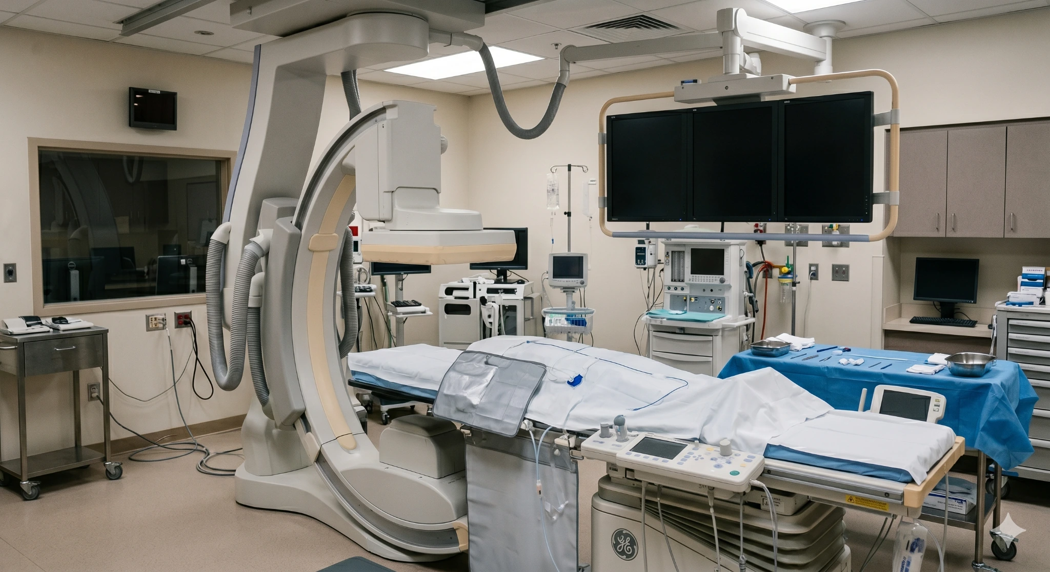
When endovascular treatment is appropriate, catheter-based tools may be guided to a blockage using live imaging. The access site, anesthesia, device choice, and recovery plan depend on the patient and the anatomy.
A small balloon is guided to the narrowing and inflated, pressing plaque outward and restoring the channel.
A specialized catheter may be used to modify or remove selected plaque before another therapy is applied.
The same balloon technique, coated with medication that discourages the artery from renarrowing afterwards.
A mesh scaffold is placed where the vessel would otherwise collapse, holding the artery open long term.
Candidacy
A procedure should be considered when it is likely to improve function, relieve persistent symptoms, or protect a threatened limb—not simply because a scan shows narrowing.
What happens
No surprises. Here is the sequence from first phone call to follow-up.
Your care team
Meet the EVSLA physicians featured in this concept. Credentials, specialties, biographies, and care-pathway claims should be matched to the practice’s approved source content before publication.

Endovascular Specialist
Treats complex arterial disease across all three clinics, with a focus on limb preservation in patients who have been told amputation is the only option.

Endovascular Specialist
Works closely with referring primary care physicians and podiatrists on early PAD detection and coordinated diabetic foot care.

Endovascular Specialist
Fellowship-trained in vascular and interventional radiology, covering arterial disease, venous conditions, and interventional oncology.
Where we treat PAD
Choose the location most convenient for you. Confirm the testing and treatment services offered at each clinic before publishing this section.
445 W Broadway
Glendale, CA 91204
2750 Sycamore Dr, Ste 200
Simi Valley, CA 93065
1407 Foothill Blvd
La Cañada, CA 91011
Questions patients ask
Go to an emergency department if a leg or foot becomes suddenly cold, pale or blue, numb, and painful — particularly if it happens over minutes to hours. That pattern can mean an artery has closed acutely, and the window to save the limb is short.
Also seek same-day care for a foot wound with spreading redness, fever, or a foul smell. Ordinary claudication that has been stable for months is not an emergency, but it does warrant a scheduled evaluation.
Advanced PAD can cause foot pain that becomes worse when the leg is raised and temporarily improves when the foot is lowered. That pattern can be a sign of severely reduced blood flow.
Do not rely on sleeping position as treatment. New or worsening rest pain, color change, coldness, numbness, or a wound that is not healing needs prompt medical assessment.
Most commonly atherosclerosis — cholesterol and calcium building into plaque along the artery wall over years. Smoking, diabetes, high blood pressure, high cholesterol, and age are the principal drivers.
Less often, leg artery blockage results from inflammation of the vessels, injury, radiation exposure, or a clot traveling from elsewhere. This distinction matters, because those causes are treated differently.
Plaque that has already formed does not usually disappear. What can change is how much blood reaches your leg and how far you can walk — through risk-factor control, a structured walking program, medication, and where appropriate a procedure to reopen the vessel.
Stopping smoking and treating cholesterol and diabetes can meaningfully slow or halt progression. Nobody should promise you reversal; a specialist should be able to tell you realistically what improvement to expect in your case.
Staying properly hydrated supports circulation generally and matters before and after procedures that use contrast dye. It is not, however, a treatment for arterial narrowing, and no amount of water will reopen a blocked vessel.
The interventions with real evidence behind them are stopping tobacco, supervised walking, statin and antiplatelet therapy, and glucose control.
Coverage for diagnostic testing and medically necessary revascularization depends on your plan, documented symptoms, test results, and network rules.
Ask the practice to confirm whether it verifies benefits and estimates patient responsibility before scheduling, then replace this sentence with the approved billing language.
Not for most plans — you are welcome to contact us directly. Some HMO plans do require a referral from your primary care physician, which our team can help you confirm.
If you were referred by a physician, we send them a full report of what we found and what we did.
A focused vascular evaluation may include a pulse exam, an ankle-brachial index, and additional imaging when appropriate. The goal is to understand whether circulation is contributing to your symptoms and what options fit your situation.
Coverage, testing, and treatment setting are confirmed individually.