


A toe or foot sore stays open
A blister, cut, pressure spot, or ulcer that remains open for two weeks can be part of CLTI when objective testing also shows poor arterial blood flow.
Limb-saving arterial care · Los Angeles
Foot pain at rest, a wound that is not healing, or darkening tissue can mean the limb is not receiving enough blood. EVSLA evaluates the blood-flow problem promptly, then explains which medical, wound-care, endovascular, or surgical pathway fits the situation.
A limb warning worth acting on
Chronic limb-threatening ischemia (CLTI), historically called critical limb ischemia, is an advanced form of peripheral artery disease. It combines objectively reduced leg blood flow with ischemic pain at rest, gangrene, or a lower-limb ulcer that has been present for more than two weeks.
CLTI is not diagnosed from a photograph or pressure number alone. The symptoms, wound, blood-flow measurements, infection status, anatomy, and overall health all matter. Suspected CLTI needs urgent specialist assessment because delayed healing can place tissue and the limb at risk.
Sources: Society for Vascular Surgery — CLTIand Global Vascular Guidelines
Warning signs
CLTI symptoms can overlap with neuropathy, infection, pressure injury, and vein disease. The combination of pain, a wound, and poor blood flow is what raises concern—so let an urgent exam and objective testing determine the cause.
Do not wait for every sign to appear. Rest pain, a wound present for two weeks, or gangrene is enough to justify urgent specialist assessment. Sudden symptoms are an emergency.
What to look for
Photos can help document change, but they cannot show how much blood reaches the foot. Photograph the wound, note when it began, and arrange prompt assessment rather than waiting for a more dramatic appearance.
A blister, cut, pressure spot, or ulcer that remains open for two weeks can be part of CLTI when objective testing also shows poor arterial blood flow.

An ischemic wound may appear dry, sharply defined, or pale at the edges. Appearance alone is not enough to identify the cause or the needed treatment.
The foot may become pale when raised and dusky red when lowered. Compare both sides and report a new or worsening difference promptly.

A persistent temperature difference may accompany reduced circulation. Sudden coldness with pain, pallor, numbness, or weakness is an emergency.
Darkening tissue can represent gangrene and needs immediate medical attention. Do not cut, soak, or treat it with home remedies.
Increasing redness, swelling, drainage, odor, fever, or chills can signal infection. In a poorly perfused foot, infection can progress quickly.
A wound can have more than one cause—arterial, venous, neuropathic, infectious, traumatic, or pressure-related. Treating the surface without checking perfusion can miss the problem underneath.
Urgent is not the same as sudden
CLTI usually develops over time, but it still needs urgent vascular assessment. A sudden loss of circulation is acute limb ischemia, a different and time-sensitive emergency. Infection can also turn an urgent wound into an emergency.
Sources: Society for Vascular Surgery PAD guide and Acute limb ischemia guide .
How clinicians assess limb threat
Treatment is not chosen from one scan or one pressure number. A specialist considers the wound, the measured loss of blood flow, infection, the arterial anatomy, and the patient’s overall health before recommending a limb-preservation plan.
W — Wound
The size, depth, location, and healing trend of an ulcer or gangrene help describe the current limb threat.
I — Ischemia
Toe pressure, toe-brachial index, ABI, waveforms, and sometimes tissue-perfusion testing quantify the circulation deficit.
fI — Foot infection
Redness, drainage, depth, systemic symptoms, and imaging can change the urgency and sequence of treatment.
PLAN — Treatment fit

Patient risk, limb severity, anatomy, available conduit, goals, and preferences shape endovascular, surgical, or combined care.
The WIfI framework stands for Wound, Ischemia, and foot Infection. It helps clinicians stage—not self-diagnose—the threatened limb. The PLAN framework adds Patient risk, Limb severity, and ANatomic complexity.
(Global Vascular Guidelines)Diagnosis
CLTI evaluation begins with the history, pulse and foot exam, and objective blood-flow testing. ABI is useful, but diabetes and calcified arteries can make ankle readings misleadingly high. Toe pressures and waveforms often add essential information.
How the diagnostic pieces fit
Global guidelines call for objective hemodynamic testing and identify toe pressures as a preferred measure in suspected CLTI. Test choice still depends on the patient and local capability. Read the guideline..
Treatment
CLTI care is coordinated, not one-dimensional. Cardiovascular risk treatment, wound and infection care, pressure offloading, and restoration of blood flow may all be needed. The sequence changes when infection or nonviable tissue is present.
Whole-patient care
CLTI is advanced PAD, so care includes proven cardiovascular protection and management of conditions that affect healing.
Local limb care
Wound care is coordinated with blood-flow evaluation. Infection and pressure can block healing even after circulation improves.
Restore flow
The goal is to establish enough in-line blood flow to relieve ischemic pain and support healing. No procedure can guarantee wound closure or limb salvage.
Endovascular options
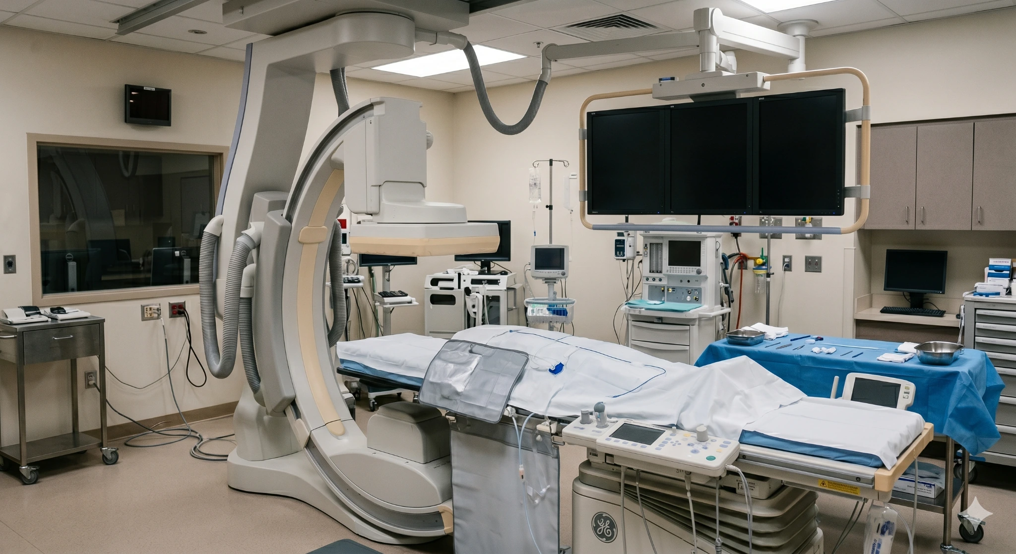
When an endovascular approach fits the limb and anatomy, catheter-based tools may be guided through the artery using live imaging. The device, access site, anesthesia, treatment setting, and recovery plan depend on the patient and the target vessel.
A small balloon is guided to a narrowed segment and inflated to enlarge the channel. The goal is improved in-line flow toward the threatened tissue.
A specialized catheter may modify or remove selected plaque when the lesion and treatment plan make that useful.
A medication-coated balloon may be considered in selected arteries to reduce renarrowing after the vessel is opened.
A mesh scaffold may be placed when the vessel needs support after treatment. Stents are not appropriate for every artery or lesion.
Candidacy
A procedure is considered when restoring blood flow is likely to relieve ischemic pain, support healing, or help preserve a functional limb. The decision is based on the whole patient—not simply a blocked artery on a scan.
What happens
CLTI care may move faster than an ordinary consultation. The order can change when infection, wet gangrene, or sudden symptoms are present.
Your care team
CLTI is best handled as a coordinated limb-preservation problem. The physicians below are included from the approved EVSLA concept; credentials, biographies, hospital relationships, and specific service claims must be confirmed before publication.

Endovascular Specialist
Featured in this concept for image-guided evaluation and treatment of complex lower-extremity arterial disease. Confirm approved limb-preservation language before publication.

Endovascular Specialist
Featured in this concept for coordinated arterial assessment with referring primary care, podiatry, and wound-care clinicians. Confirm the care pathway before publication.

Endovascular Specialist
Featured in this concept for vascular and interventional radiology care. Confirm training, specialty description, and CLTI services against approved source content.
Where to request a CLTI evaluation
Call before traveling so the team can triage urgency and direct you to the right setting. Confirm CLTI testing, treatment capability, scheduling, and hospital relationships for each location before publication.
445 W Broadway
Glendale, CA 91204
2750 Sycamore Dr, Ste 200
Simi Valley, CA 93065
1407 Foothill Blvd
La Cañada, CA 91011
Questions patients ask
CLTI is an urgent condition that needs prompt specialist assessment. It usually develops over time, with ischemic rest pain, a non-healing wound, or gangrene.
Call 911 or go to an emergency department if symptoms begin suddenly—especially severe pain, a cold or pale limb, numbness, weakness, or inability to move the foot. Also seek immediate care for rapidly spreading infection, wet gangrene, fever, or systemic illness.
The terms are often used for the same advanced PAD syndrome. Chronic limb-threatening ischemia is now preferred because limb risk exists on a continuum and cannot be defined by one pressure threshold alone.
Many patients and websites still use “critical limb ischemia,” so this page includes both terms. The diagnosis requires symptoms or tissue loss plus objective evidence of poor arterial blood flow.
Ischemic rest pain is often described as burning, aching, or severe discomfort in the forefoot or toes when a person is lying down. It may worsen when the foot is raised in bed and temporarily improve when the foot hangs down.
Neuropathy, arthritis, infection, and other conditions can cause pain at night too. The pattern is a warning sign, not a diagnosis; it needs an exam and blood-flow testing.
Yes. Healing requires oxygen and nutrients delivered by the bloodstream. A cut, blister, pressure injury, or diabetic foot ulcer may remain open when arterial flow is too low.
Not every non-healing wound is arterial. Venous disease, neuropathy, pressure, trauma, infection, and inflammation can contribute, sometimes together. That is why the wound and circulation should be evaluated together.
Often the treatment goal is limb preservation through coordinated medical care, wound and infection treatment, pressure offloading, and revascularization when appropriate. Options may include an endovascular procedure, surgical bypass, or a hybrid approach.
No clinician can promise limb salvage. When tissue is nonviable, infection is uncontrolled, the limb cannot be reconstructed, or the expected burden outweighs the benefit, amputation may be the safest option. That decision should follow multidisciplinary assessment and shared decision-making whenever circumstances allow.
Diagnosis combines the clinical problem—rest pain, a wound, or gangrene—with objective evidence of PAD. Testing may include ABI, toe pressure or toe-brachial index, arterial waveforms, duplex ultrasound, and additional perfusion testing.
If revascularization is being considered, CTA, MRA, or catheter angiography may be used to map the arterial anatomy. In diabetes or chronic kidney disease, ankle vessels can be noncompressible, which is one reason toe measurements are important.
Most plans include cardiovascular risk treatment, wound care, infection management when needed, pressure offloading, and preventive foot care. Revascularization may be recommended to restore blood flow and can be endovascular, surgical, or hybrid.
The best approach depends on the patient’s procedural risk, the severity of limb threat, the anatomy, availability of a suitable vein for bypass, prior treatments, and the patient’s preferences. Treatment remains individualized even when two wounds look similar.
Elevation can make ischemic foot pain worse because gravity is no longer helping blood reach the foot. Some people temporarily lower the foot for relief, but positioning does not treat the blocked artery and can increase swelling.
Do not use sleep position as a substitute for care. New or worsening rest pain, color change, coldness, numbness, weakness, or a wound that is not healing needs prompt assessment.
Coverage for diagnostic testing, wound care, and medically necessary revascularization depends on the plan, documented symptoms, objective findings, network rules, and the proposed setting.
Before publication, replace this concept language with EVSLA’s approved benefit-verification and financial-responsibility policy.
A CLTI evaluation may include a wound and pulse exam, ABI and toe pressures, arterial waveforms, and anatomic imaging when appropriate. The goal is to identify what is preventing healing, how urgent the limb threat is, and which coordinated care path fits the patient.
For sudden severe pain, coldness, numbness, weakness, or color loss, call 911.