
Aorta · abdomen

Abdominal aortic aneurysm
An AAA forms in the aorta below the chest, often below the kidney arteries. Ultrasound can screen and monitor many AAAs; CTA maps anatomy when repair is considered.
Aneurysm evaluation · Los Angeles
An aneurysm is a weakened artery wall that has enlarged. It may grow without warning signs. EVSLA reviews the location, diameter, shape, growth, anatomy, and whole-patient risk to explain whether surveillance, endovascular repair, open surgery, or specialty referral makes sense.
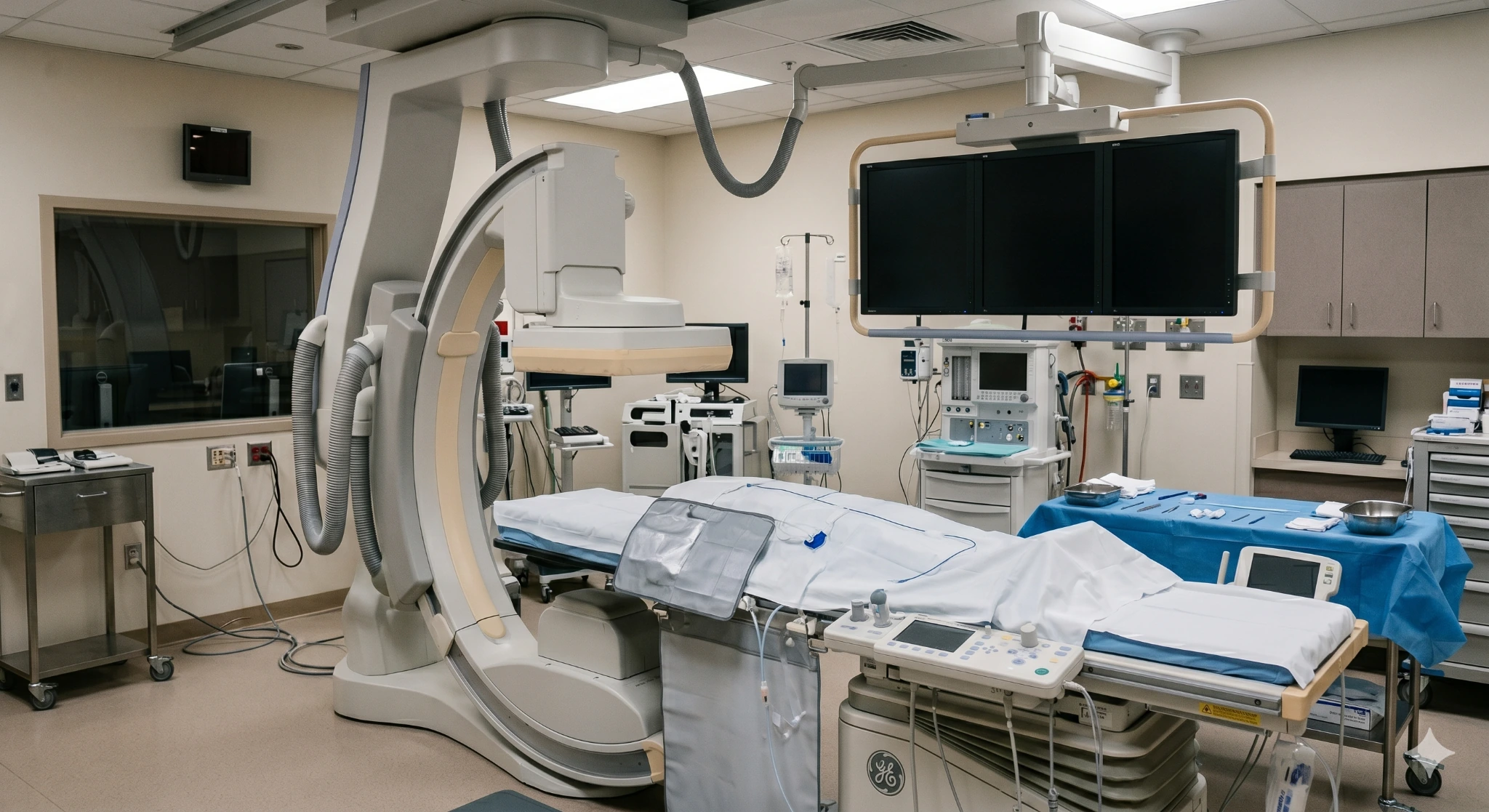
A finding worth reviewing
An aneurysm is an abnormal enlargement of an artery caused by weakening of the vessel wall. The aorta is the body’s largest artery; aneurysms can also form in iliac arteries in the pelvis or peripheral arteries such as the popliteal artery behind the knee.
Many aneurysms cause no symptoms and are discovered on imaging. The important questions are where the aneurysm is, how large it is, whether it is growing or causing symptoms, and whether its anatomy makes surveillance, endovascular repair, open repair, or multidisciplinary referral the safest path.
Sources: Society for Vascular Surgery — Abdominal Aortic Aneurysms and 2022 ACC/AHA Aortic Disease Guideline
Location changes the problem
“Aneurysm” describes the vessel-wall problem, not a single treatment. The artery involved determines the imaging, rupture or clotting risk, repair options, and specialist team.
Aorta · abdomen
An AAA forms in the aorta below the chest, often below the kidney arteries. Ultrasound can screen and monitor many AAAs; CTA maps anatomy when repair is considered.
Pelvis

An aneurysm in a pelvic artery may occur alone or with an AAA. Branch anatomy matters when planning an endovascular graft or open repair.
Behind the knee

This peripheral aneurysm can form clot, send debris to the foot, or suddenly block leg blood flow. Duplex ultrasound is central to evaluation and follow-up.
Aorta · chest

Aneurysms in the chest, aortic root, or ascending aorta require location-specific cardiovascular or cardiothoracic expertise and a multidisciplinary aortic team.
An aneurysm is not the same as an aortic dissection. An aneurysm is enlargement of the vessel; a dissection is a tear within the aortic wall. Both can become emergencies, but the workup and treatment are not interchangeable.
(ACC/AHA Guideline)Possible warning signs
These symptoms can have many causes and do not diagnose an aneurysm. A known aneurysm with new pain deserves prompt medical advice; sudden severe symptoms need emergency care.
Do not press repeatedly on a suspected pulsatile mass. A clinician should determine whether the finding is vascular and select the appropriate imaging.
Emergency symptoms
An aneurysm may rupture, tear, clot, or send embolic debris downstream. Those complications can become life- or limb-threatening within minutes.
Sources: SVS — Patients with AAA and SVS Popliteal Aneurysm Guideline .
Risk and screening
AAA risk rises with age, tobacco exposure, family history, high blood pressure, atherosclerotic disease, and aneurysm in another artery. A person with one aneurysm may be evaluated for aneurysms elsewhere.
Screening guidance applies to people without symptoms. New pain or emergency symptoms need diagnostic evaluation, not routine screening. Source: USPSTF — Abdominal Aortic Aneurysm Screening .
Diagnosis and surveillance
One scan gives a diameter. The care plan also needs location, morphology, branch-vessel anatomy, symptoms, prior measurements, and the clinical context.
Five questions guide the workup
The ACC/AHA guideline emphasizes consistent imaging acquisition, measurement, and reporting. Read the guideline summary..
Treatment decision
Repair is recommended when the predicted risk of rupture, thrombosis, embolization, or other complication outweighs procedural risk. The balance is individualized.
Monitor
Smaller, stable, asymptomatic aneurysms are often followed with scheduled imaging while cardiovascular risks are addressed.
Repair from within
EVAR or a covered stent-graft may exclude selected aneurysms from circulation through catheter-based access.
Reconstruct
Open surgery may offer the better durable option for some patients or anatomies. Root and ascending aortic disease usually requires a cardiothoracic pathway.
Endovascular repair
EVAR places a stent-graft inside the artery so blood travels through the graft instead of pressurizing the aneurysm sac. It is less invasive than open abdominal repair, but it is not minor and it is not appropriate for every anatomy.
CTA measurements define the aneurysm, access arteries, seal zones, angulation, kidney arteries, and pelvic branches.
Catheters and the graft system are advanced through access arteries, commonly from the groin, under live X-ray guidance.
The stent-graft is positioned to channel blood past the aneurysm while preserving critical branches whenever possible.
Follow-up imaging checks graft position, aneurysm-sac size, blood flow, and endoleak. Surveillance remains necessary even when recovery is smooth.
Endovascular popliteal or iliac aneurysm repair uses different devices and anatomic criteria. Some patients are better served by open bypass or reconstruction. The repair must match the artery, the downstream circulation, and the patient’s expected durability needs.
Repair candidacy
Aneurysm size is important, but candidacy is a balance of complication risk, anatomy, procedural risk, life expectancy, and patient goals.
What happens
Bring the actual imaging disc or portal access when possible—not only the report. Earlier studies are especially valuable for measuring growth.
Your care team
Aneurysm care often crosses specialties. EVSLA can evaluate the vascular and endovascular component and coordinate referral when open vascular, cardiothoracic, genetic-aortopathy, or hospital-based care is the safer pathway. Physician credentials and exact aneurysm services must be confirmed before publication.

Endovascular Specialist
Featured in this concept for image-guided vascular evaluation and endovascular planning. Confirm approved AAA, EVAR, iliac, and peripheral aneurysm service language before publication.

Endovascular Specialist
Featured in this concept for arterial imaging review, endovascular candidacy, and coordination with vascular or cardiothoracic surgery. Confirm the actual pathway before publication.
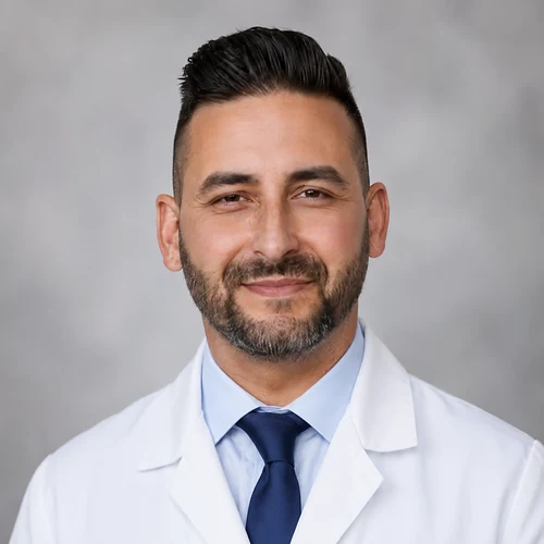
Endovascular Specialist
Featured in this concept for vascular and interventional radiology care. Confirm training, specialty description, repair capability, and hospital relationships against approved content.
Where to request an aneurysm evaluation
Call before traveling so the team can review the referral, identify any emergency concern, and confirm the correct clinic or hospital pathway. Verify imaging access, EVAR capability, surgical partnerships, and scheduling at each location before publication.
445 W Broadway
Glendale, CA 91204
2750 Sycamore Dr, Ste 200
Simi Valley, CA 93065
1407 Foothill Blvd
La Cañada, CA 91011
Questions patients ask
An aneurysm is abnormal enlargement caused by weakening of an artery wall. An aortic dissection is a tear in the inner aortic wall that allows blood to separate layers of the vessel.
They can occur separately or together, and both can become emergencies. Sudden severe chest, back, or abdominal pain, fainting, or neurologic symptoms requires emergency care.
Yes. Many abdominal and thoracic aortic aneurysms are discovered incidentally during imaging for another reason or through screening. Lack of pain does not show whether an aneurysm is stable.
A known aneurysm needs a defined surveillance plan. The interval depends on its location, size, growth, anatomy, and the guideline used by the treating team.
There is no single number for every patient. The Society for Vascular Surgery recommends elective repair for many patients at acceptable risk with a typical fusiform AAA at or above 5.5 cm, and suggests repair for women with AAA between 5.0 and 5.4 cm.
Symptoms, saccular shape, concerning growth, body size, anatomy, sex, operative risk, and patient preference can change timing. Do not use a report measurement to make this decision without specialist review.
The USPSTF recommends one-time ultrasound screening for men age 65 to 75 who have ever smoked and selective screening for men in that age group who have never smoked.
For women, recommendations depend on smoking and family history, and evidence is less certain in some groups. Screening guidance is for people without symptoms; symptoms need diagnostic evaluation.
Endovascular aneurysm repair uses a stent-graft delivered through arteries, commonly from the groin, to create a new channel inside the aorta and reduce pressure on the aneurysm wall.
EVAR requires suitable anatomy and careful graft sizing. It usually avoids a large abdominal incision but still carries risks and requires lifelong imaging for endoleak, graft position, and aneurysm-sac change.
Neither approach is best for everyone. EVAR is less invasive and often has a shorter initial recovery, while open repair may offer the more appropriate or durable solution for certain anatomies and patients.
The choice considers age, overall health, life expectancy, aneurysm anatomy, access vessels, kidney function, prior surgery, ability to complete surveillance, and the patient’s priorities.
Yes. A popliteal aneurysm can form clot, suddenly thrombose, or send embolic material into smaller arteries of the foot. Symptoms can include a cold painful foot, blue toes, numbness, weakness, or a non-healing wound.
Sudden symptoms can indicate acute limb ischemia and require emergency assessment. Stable aneurysms still need surveillance or timely repair planning.
The interval depends on location, diameter, growth, symptoms, imaging quality, and whether the aneurysm has been repaired. Smaller stable aneurysms are generally scanned less often than larger or changing aneurysms.
After EVAR, imaging begins early and continues long term. Follow the schedule from the treating team rather than applying another person’s interval to your scan.
There is no medication that reliably reverses an established degenerative aneurysm. Medical care focuses on smoking cessation, blood-pressure and cardiovascular risk management, and treatment of related disease.
Do not start or stop medication based on general website information. The prescribing clinician should tailor treatment to the patient’s full cardiovascular and medical history.
Call 911 for sudden severe chest, back, abdominal, flank, groin, or leg pain; fainting; clammy weakness; difficulty breathing; new neurologic symptoms; or a suddenly cold, pale, numb, weak, or immobile limb.
Do not drive yourself. Tell emergency personnel if you have a known aneurysm and where it is located.
Bring the imaging report, source images, and any earlier scans. EVSLA can review the vascular anatomy, explain surveillance and endovascular considerations, and coordinate a surgical or multidisciplinary referral when that is the safer pathway.
For sudden severe pain, fainting, or sudden loss of limb circulation, call 911.


