
Tiny surface vessels

Spider veins
Fine red, blue, or purple webs close to the skin. Often cosmetic, but some itch, burn, or bleed.
Visible vein care · Los Angeles
Bulging, twisted, blue, red, or web-like veins can be cosmetic—but aching, heaviness, swelling, itching, skin changes, or bleeding may point to a pressure problem underneath. EVSLA evaluates the symptom pattern and, when indicated, maps reflux with duplex ultrasound before matching treatment to the actual vein.
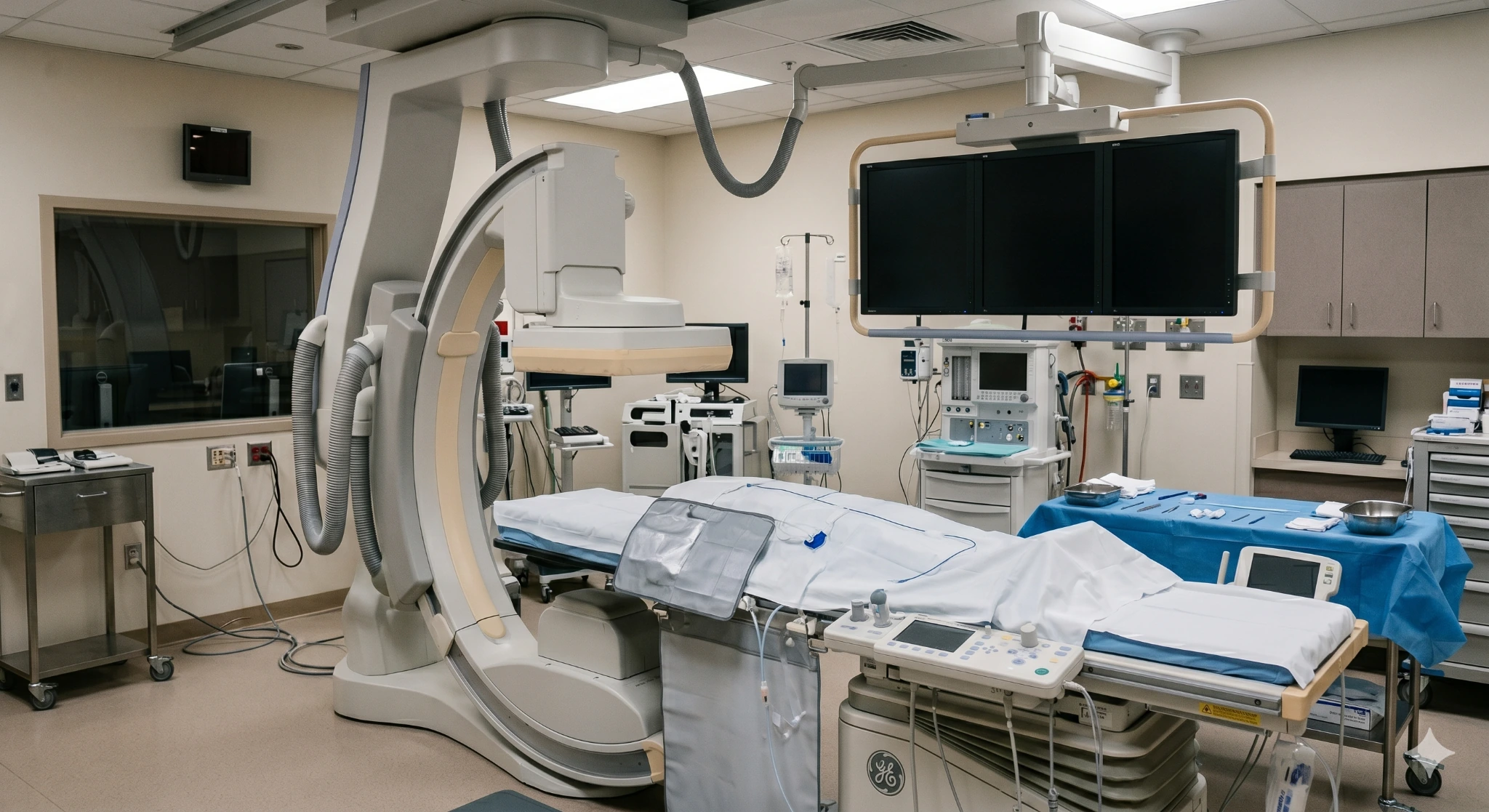
A pattern worth mapping
Varicose veins are enlarged, twisted surface veins that often bulge above the skin. They commonly form when one-way valves stop closing effectively and blood moves backward—called reflux—raising pressure in connected branches. They may ache, feel heavy, itch, swell, inflame, bleed, or contribute to skin changes.
Spider veins are much smaller red, blue, or purple vessels close to the skin. Many are primarily cosmetic, although they can itch, burn, or bleed and may coexist with deeper reflux. Appearance alone cannot show which vein is feeding the pattern or whether treatment should begin deeper.
Clinical references: NHLBI and Society for Vascular Surgery and NICE guidance
Spider veins vs. varicose veins
Vein size and appearance guide the exam, but symptoms and ultrasound findings determine whether the goal is cosmetic improvement, pressure relief, or treatment of an underlying reflux source.
Tiny surface vessels
Fine red, blue, or purple webs close to the skin. Often cosmetic, but some itch, burn, or bleed.
Feeder veins

Blue-green veins beneath the skin that may feed clusters of spider veins or connect to larger branches.
Bulging branches

Raised, twisted veins that may ache, feel heavy, swell, itch, inflame, or bleed.
Pressure complication

Brown staining, eczema, firmness, fragile skin, or an ankle sore suggests more advanced venous disease.
Appearance guides the exam; symptoms and duplex findings guide medical treatment.
American Venous Forum guidelinesVaricose vein symptoms
Symptoms often build after long periods of standing or sitting and may improve with walking, elevation, or rest. The same complaints can come from other conditions, so the pattern should be assessed rather than assumed.
What to photograph
Take pictures in the same lighting and note whether the pattern changes after standing, exercise, heat, elevation, pregnancy, or prior treatment. Images support the history; they do not replace an exam or duplex.
Small spider veins often cluster around the thigh, knee, ankle, or foot.
Reticular veins sit below the skin and may supply smaller visible vessels.
Bulging veins may become more prominent while standing or later in the day.

Indentation around the ankle can add evidence of venous pressure or another swelling cause.
Skin changes near the ankle make the problem more than cosmetic and deserve assessment.

A wound, recurrent bleeding site, or sore near a vein should be evaluated promptly.
When to worry about varicose veins
Most visible veins are not emergencies. A sudden or rapidly changing symptom can point to bleeding, superficial thrombophlebitis, DVT, infection, an ulcer, or an arterial problem.
Call 911 for sudden shortness of breath, chest pain—especially with breathing—coughing blood, fainting, or severe weakness.
Clinical reference: NHLBI, , .
What causes varicose and spider veins?
Visible veins develop through more than one pathway. Treatment planning asks whether the problem is limited to small surface vessels, connected to superficial reflux, or influenced by deeper obstruction or prior clot damage.
Mechanism
Blood falls backward while upright, raising pressure in a saphenous trunk or connected branches and stretching the vein wall.
Predisposition
Inherited vein-wall traits, hormonal changes, increased blood volume, and pregnancy pressure can make visible veins more likely.
Contributors
Prolonged standing or sitting, limited calf movement, extra body weight, and prior DVT can increase pressure or worsen symptoms.
Diagnosis before treatment
The evaluation compares appearance with symptoms, standing exam findings, prior pregnancy and clot history, skin changes, pulses, and previous treatment. Duplex ultrasound is used when the pattern suggests truncal reflux, recurrence, deep-vein disease, or a procedure that depends on anatomy.
Tiny spider veins without symptoms may be assessed as a surface treatment problem. A symptomatic varicose branch should not be injected or removed until the clinician decides whether a feeding reflux source must be treated first.
What a visible-vein evaluation checks
Varicose and spider vein treatment
Not every visible vein needs treatment. When symptoms, complications, or appearance goals justify care, the sequence is based on duplex findings, vein size, location, skin health, prior procedures, and patient preferences.
Support symptoms
Position changes and calf movement can reduce pooling. Clinician-selected compression may ease aching, heaviness, and swelling but does not repair a leaking valve.
Close the reflux source
When a refluxing truncal vein feeds symptomatic branches, thermal or nonthermal closure may redirect flow through functioning veins.
Treat visible branches
After the source is addressed—or when no truncal reflux is present—selected spider veins, feeder veins, and bulging branches can be treated directly.
Procedures we may discuss
The procedure name should follow the anatomy—not lead it. A spider-vein injection, bulging-branch removal, and saphenous-vein closure solve different parts of the problem.

Controlled heat closes a selected refluxing truncal vein through an ultrasound-guided catheter.
Laser energy closes a refluxing saphenous vein through a small catheter placed with ultrasound guidance.
Liquid or foam medication seals selected spider, reticular, or varicose veins so they fade over time.
Bulging surface branches are removed through very small openings after local numbing.
Recovery and results
Recovery depends on the procedure, number and size of treated veins, medications, skin health, and work demands. The treating clinician should give the actual schedule.
First days

Many patients walk soon after office-based treatment. Compression and activity guidance vary by procedure.
Early healing
Bruising, tenderness, tightness, firmness, or discoloration can occur while treated veins close and resolve.
Weeks to months
Spider veins may need staged sessions. Larger branches can take time to soften and become less visible.
Long term
Treatment closes selected veins; it does not remove inherited risk or guarantee that no new visible veins form.
Recovery and the pace of visible improvement vary by procedure and treatment area.
NHLBI treatment overviewWho should be evaluated
A vein evaluation is reasonable when symptoms persist, daily activity is affected, veins recur after treatment, or skin and bleeding changes make the issue more than cosmetic.
What happens
Bring symptom timing, pregnancy and clot history, prior ultrasound reports, compression garments, medication list, and photographs of changes or bleeding.
Your care team
This concept positions EVSLA’s physicians around symptom review, duplex interpretation, reflux treatment, and coordination when the pattern suggests DVT, lymphedema, pelvic obstruction, wound-care needs, or another specialty. Physician credentials and exact services must be confirmed before publication.
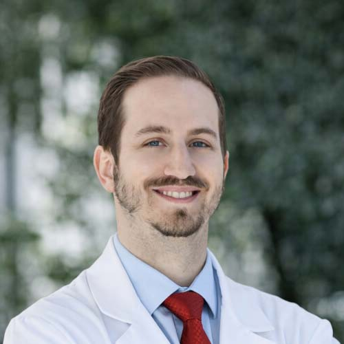
Endovascular Specialist
Featured in this concept for venous imaging review and minimally invasive treatment planning. Confirm approved credentials and procedure scope before publication.

Endovascular Specialist
Featured in this concept for reflux mapping, treatment candidacy, and image-guided venous care. Confirm services and location availability.
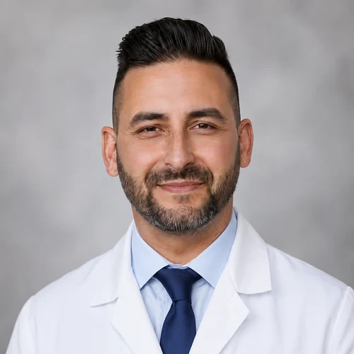
Endovascular Specialist
Featured in this concept for ultrasound-led assessment and treatment planning. Confirm training, specialty description, and approved claims.
Where to request a vein evaluation
Call before traveling to confirm that diagnostic duplex and the recommended visible-vein treatment are available at the selected clinic. Exact services and scheduling must be verified before publication.
445 W Broadway
Glendale, CA 91204
2750 Sycamore Dr, Ste 200
Simi Valley, CA 93065
1407 Foothill Blvd
La Cañada, CA 91011
Questions patients ask
Varicose veins usually form when vein walls stretch and one-way valves do not close effectively. Blood can flow backward and pool, raising pressure and enlarging connected surface branches.
Family history, age, pregnancy, long periods of standing or sitting, limited calf movement, extra body weight, and prior clot damage can increase risk or worsen the pattern.
Spider veins are fine red, blue, or purple vessels close to the skin. Varicose veins are larger, twisted branches that often bulge above the skin and are more likely to cause aching, heaviness, swelling, itching, inflammation, or bleeding.
Both can coexist. Symptoms and duplex findings—not color alone—help determine whether treatment should begin with an underlying reflux source.
Arrange prompt assessment for active or recurrent bleeding, a hot red tender cord, sudden one-sided swelling, rapidly worsening pain, skin darkening, spreading redness, or an open sore.
Call 911 for sudden chest pain, shortness of breath, coughing blood, fainting, or severe weakness because pulmonary embolism is possible.
Most spider veins are primarily an appearance concern and are not dangerous. Some itch, burn, or bleed, and a widespread pattern may coexist with reticular veins or deeper reflux.
New symptoms, one-sided swelling, skin changes, recurrent bleeding, or a wound should not be dismissed as cosmetic.
The best treatment depends on the source and goal. Confirmed reflux in a symptomatic truncal vein may be treated with radiofrequency ablation, laser ablation, or another closure method. Bulging branches may need microphlebectomy or sclerotherapy.
Duplex mapping helps avoid treating only the visible branch while leaving the feeding reflux untreated.
Liquid sclerotherapy is commonly used for spider veins on the legs. A clinician injects a medication that seals selected vessels so they gradually fade. Some patterns require more than one session.
Feeder veins or underlying reflux may need to be addressed first. Surface laser is used in some settings, especially for very small vessels, but candidacy depends on the pattern and skin.
Compression can reduce pooling, aching, heaviness, and swelling, but it does not repair a damaged valve or permanently remove a varicose vein.
The strength and fit should be individualized. People with a cold painful foot, possible arterial disease, severe neuropathy, or certain heart conditions should seek guidance before strong compression.
Topical creams may moisturize skin or temporarily soothe irritation, but they do not close a refluxing vein or remove established spider veins. Exercise, position changes, elevation, and healthy weight can help symptoms and venous function but do not erase every visible vessel.
A medical evaluation is appropriate before relying on a product marketed as a permanent cure.
Treated veins can close successfully, but venous disease is chronic and new visible veins may develop over time. Residual feeder veins, progression in other segments, pregnancy, and inherited risk can contribute.
Follow-up and a staged plan help distinguish normal residual vessels from recurrence that needs another duplex study.
Cost depends on the diagnosis, procedure, number of veins or sessions, imaging, location, and insurance rules. Treatment for symptomatic reflux or complications may be handled differently from cosmetic spider-vein care.
Ask for a written estimate after the evaluation identifies the actual treatment sequence. Coverage is plan-specific and should be verified directly.
Bring photographs, symptom timing, prior ultrasound reports, pregnancy and clot history, compression garments, and previous treatment records. EVSLA can separate appearance from underlying reflux and explain the right sequence.
For active bleeding that will not stop, sudden chest symptoms, or severe shortness of breath, seek emergency care.

