
Anatomy can be incidental
A compressed-looking vein on a scan does not prove that it is causing symptoms.
Pelvic venous outflow · Los Angeles
Persistent swelling, heaviness, pain, or visible veins in one—often left—leg can happen when the left common iliac vein is compressed in the pelvis. Because DVT and other conditions can look similar, EVSLA checks for clot and maps venous outflow before discussing treatment.
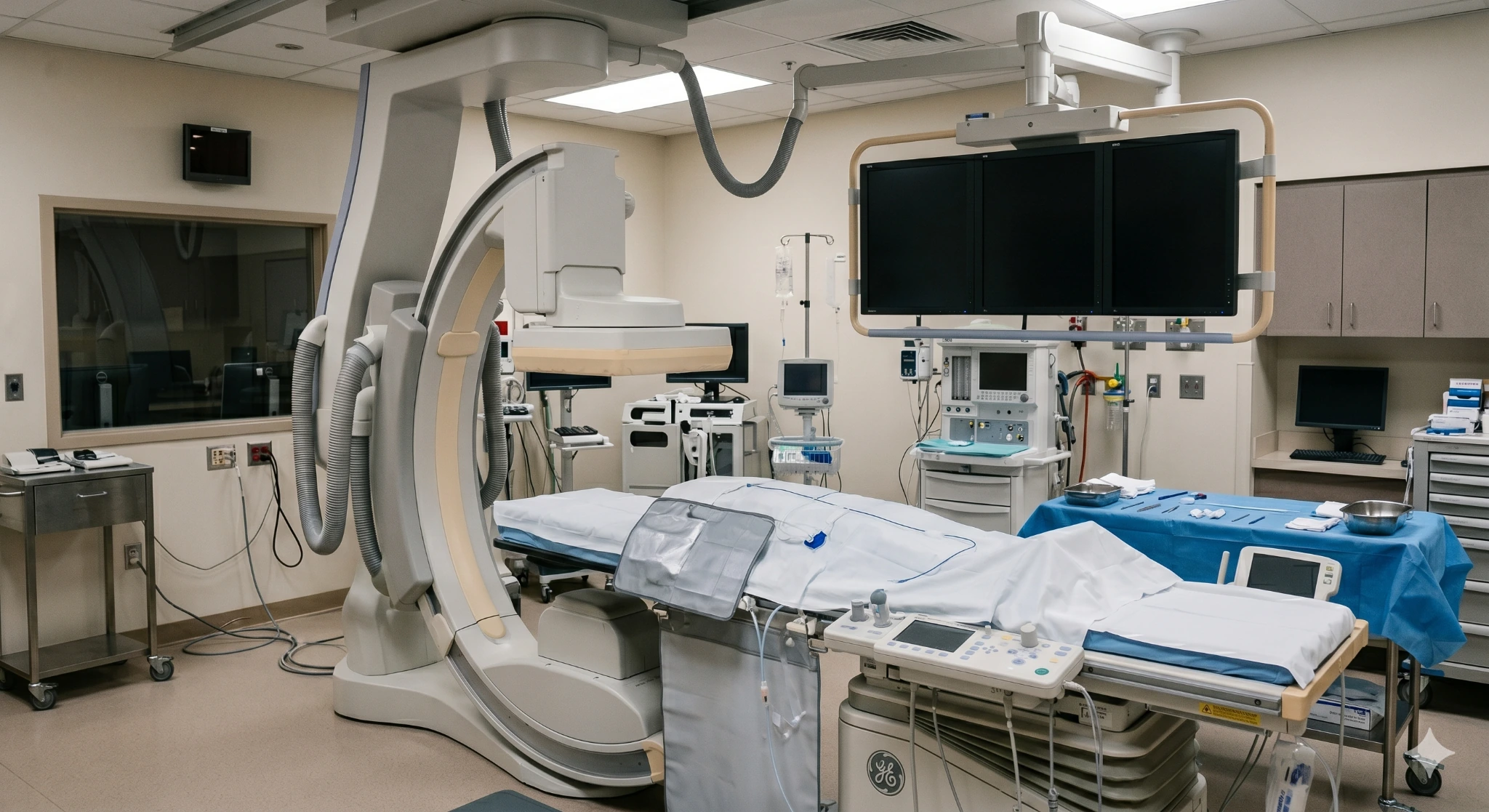
A clot warning worth checking
May-Thurner syndrome, also called iliac vein compression syndrome, is a symptomatic pelvic venous outflow problem. In the classic pattern, the right common iliac artery presses the left common iliac vein against the spine. The narrowed vein may make it harder for blood to leave the left leg, contributing to chronic swelling, heaviness, pain, varicose veins, skin changes, or deep vein thrombosis (DVT).
Many people have some iliac-vein compression on imaging without symptoms. That anatomy alone does not automatically require a stent. A useful diagnosis connects symptoms and clot history with the location and significance of the obstruction while excluding other causes.
Clinical references: Society for Vascular Surgery and 2024 multisociety NIVL consensus
The anatomy in plain language
The left common iliac vein carries blood from the left leg toward the heart. In the classic May-Thurner configuration, it passes between the right common iliac artery and the lumbar spine. Repeated arterial pulsation and limited space may narrow the vein and encourage collateral veins or clotting in susceptible patients.
A compressed-looking vein on a scan does not prove that it is causing symptoms.
Other iliac-vein compression patterns can occur, including right-sided or bilateral obstruction.
Acute DVT, old clot damage, and nonthrombotic compression are evaluated differently.
Clinical reference: Society for Vascular Surgery.
A simplified orientation—not a diagnostic image.
May-Thurner syndrome symptoms
Symptoms may be mild, build through the day, worsen with prolonged standing or exercise, or appear suddenly if a DVT forms. One symptom cannot diagnose iliac-vein compression.
Same symptom, different cause
May-Thurner syndrome is one possibility. The evaluation also looks for conditions that require a different or more urgent plan.

A new deep-vein clot can cause sudden swelling, pain, warmth, and discoloration.

Valve failure closer to the skin can cause varicose veins, aching, and ankle swelling.

Old clot damage may leave chronic obstruction, reflux, swelling, skin changes, or ulcers.

Lymphatic fluid buildup has a different exam pattern and may need compression-focused care.

Spreading redness, fever, severe tenderness, or drainage needs prompt clinical assessment.

Joint injury, medication, heart, kidney, and other conditions can also contribute to swelling or pain.
DVT and pulmonary embolism warning signs
Call a clinician promptly for new one-sided leg swelling, pain, warmth, or discoloration. Call 911 for symptoms that could mean a pulmonary embolism, including sudden shortness of breath, chest pain, fainting, a fast or irregular heartbeat, or coughing blood.
Clinical reference: American Heart Association, , .
The reason to classify the pattern
The anatomy can be silent, cause chronic symptoms, contribute to an acute clot, or coexist with damage left by an old clot. The category changes the urgency, tests, and treatment discussion.
Anatomy only
Imaging shows narrowing, but there are no meaningful leg or pelvic venous symptoms.
Chronic symptoms
Persistent one-leg swelling, heaviness, venous claudication, varices, or skin changes without acute DVT.
Acute clot

Sudden swelling and pain may reflect a deep clot extending through the pelvis.
After a prior clot
Scarred or narrowed veins may cause lasting swelling, heaviness, skin change, or ulcer risk.
Patient selection should be symptom-driven and should integrate clinical and imaging findings.
2024 multisociety NIVL consensusWhy symptoms can surface
The crossing-vessel anatomy may exist for years. Symptoms or DVT can emerge when additional factors slow flow or increase clot risk. Not every patient has an identifiable trigger.
Clot history
Old clot damage can scar the iliac vein or damage valves, creating a post-thrombotic pattern rather than a purely nonthrombotic lesion.
Temporary risk
Changes in venous flow and clot risk can make a previously silent anatomy clinically important.
Whole-patient review
Active cancer, inflammation, clotting disorders, pelvic procedures, radiation, or a mass can alter the differential and treatment plan.
May-Thurner syndrome diagnosis
The evaluation begins with history and examination, then chooses the least invasive test that can answer the next question. Pelvic outflow is harder to see than the lower leg, so no single test fits every patient.
Treatment is considered only after symptom severity, alternatives, imaging, and patient goals are aligned.
May-Thurner diagnostic pathway
Clinical references: Society for Vascular Surgery.
May-Thurner syndrome treatment
A stent is not preventive treatment for every compressed-looking vein. The plan depends on symptoms, DVT, chronic scarring, bleeding risk, anatomy, previous care, and how much the condition affects daily life.
Path 1 · Observe or support
When anatomy is incidental or symptoms are limited, the plan may focus on observation, movement, symptom management, and follow-up.
Path 2 · Treat the clot
Anticoagulation is central. Selected patients may be considered for catheter-based clot removal after symptom duration, extent, bleeding risk, and goals are reviewed.
Path 3 · Restore outflow
For carefully selected chronic nonthrombotic or post-thrombotic obstruction, a dedicated venous stent may be used to support the iliac vein.
Procedures that may enter the plan
Not every patient needs every step. The exact combination depends on whether the problem is nonthrombotic compression, acute DVT, chronic clot damage, or another diagnosis.
Contrast imaging and ultrasound from inside the vein can define the channel, reference size, and landing zones during a selected procedure.
Selected acute iliofemoral DVT may be treated with catheter-based clot removal when potential benefit outweighs bleeding and procedural risk.
A self-expanding venous stent may support a clinically significant iliac obstruction after the anatomy and symptom relationship are confirmed.

Follow-up imaging can assess stent flow, recurrent obstruction, residual clot, or another reason for persistent symptoms.
After iliac vein treatment
Timelines vary with acute clot, procedure type, access site, medications, other health conditions, and work demands. The treating clinician should provide the actual schedule.
First days
Walking, hydration, access-site care, compression, lifting, and return-to-work advice are individualized.
Medication safety
Anticoagulant or antiplatelet decisions depend on clot history, procedure, and bleeding risk. Do not stop medication without guidance.
Scheduled surveillance
Visits compare swelling, pain, function, skin, and imaging rather than relying on appearance alone.
Long term
New one-sided swelling, worsening pain, chest symptoms, or bleeding concerns should be reported promptly.
Postprocedure medication and surveillance should be individualized to clot history, procedure, and bleeding risk.
2024 multisociety NIVL consensusWho should be evaluated
The strongest signal is a persistent or recurrent one-sided venous pattern that has not been explained by a lower-leg study alone.
What happens
Bring prior imaging discs and reports, clot records, medication list, procedure notes, compression garments, pregnancy history, and photographs of changing swelling or skin.
Your care team
This concept positions EVSLA’s physicians around venous imaging review, DVT assessment, candidacy for image-guided treatment, and postprocedure surveillance. Physician credentials, exact services, and location availability must be confirmed before publication.

Endovascular Specialist
Featured in this concept for pelvic venous imaging review and minimally invasive treatment planning. Confirm approved credentials and scope before publication.

Endovascular Specialist
Featured in this concept for DVT and iliac-outflow assessment, procedure candidacy, and image-guided venous care. Confirm current services.

Endovascular Specialist
Featured in this concept for ultrasound-led assessment and venous treatment planning. Confirm training, specialty description, and approved claims.
Where to request a vein evaluation
Call before traveling to confirm where venous duplex, pelvic venous consultation, DVT treatment, venography, IVUS, or iliac vein stenting is performed. Services may differ by location.
445 W Broadway
Glendale, CA 91204
2750 Sycamore Dr, Ste 200
Simi Valley, CA 93065
1407 Foothill Blvd
La Cañada, CA 91011
Questions patients ask
May-Thurner syndrome is a symptomatic iliac-vein outflow problem. In the classic anatomy, the right common iliac artery crosses over and compresses the left common iliac vein against the spine.
Some compression is common in people without symptoms. A diagnosis should connect symptoms or DVT with meaningful obstruction and exclude other causes.
Possible early signs include persistent swelling in one leg, heaviness, aching, pressure, visible veins, or discomfort that builds with standing or activity. The left leg is classic.
Sudden one-leg swelling, warmth, pain, tenderness, or discoloration may be DVT and needs prompt assessment.
The classic crossing pattern places the left common iliac vein between the right common iliac artery and the spine. That can narrow the channel carrying blood out of the left leg.
Other pelvic vein compression patterns can affect the right side or both sides, so laterality is a clue rather than an absolute rule.
The core issue is an anatomic relationship among the iliac vein, crossing artery, and spine. Why one person becomes symptomatic while another does not is not fully explained.
It is not usually described as a straightforward inherited disorder. Family clot history can still matter because separate clotting risks may affect DVT risk.
Duplex ultrasound is useful for checking DVT, old clot changes, reflux, and flow clues, but the pelvic iliac veins can be difficult to see completely from the surface.
CT or MR venography may define pelvic anatomy. Venography and intravascular ultrasound may be used during a selected invasive evaluation.
No. People with incidental compression and no meaningful symptoms generally do not need a prophylactic stent. Mild symptoms may be managed conservatively.
Acute DVT requires a clot-treatment plan. A venous stent may be considered for selected patients with significant symptoms and confirmed iliac outflow obstruction.
Potential problems include bleeding or bruising at the access site, medication-related bleeding, stent thrombosis or restenosis, migration or placement issues, injury to nearby structures, contrast or kidney concerns, and persistent symptoms if another condition is contributing.
Many patients walk soon after an uncomplicated endovascular procedure, but return to driving, work, exercise, lifting, compression, and travel varies with the access site, clot treatment, medications, and overall health.
Follow the treating clinician’s schedule and attend surveillance imaging.
Pregnancy does not create the classic crossing anatomy, but pregnancy and the postpartum period can change venous flow and increase clot risk. Symptoms or a prior DVT should be reviewed with the obstetric and vascular teams.
No sleeping position can remove iliac-vein compression or treat a DVT. A comfortable position and modest leg elevation may help some people’s swelling, but symptom relief does not establish the diagnosis.
Do not rely on sleep position for new or worsening one-leg swelling, pain, warmth, or color change.
Bring prior ultrasound, CT, or MR images and reports; DVT and procedure records; medication list; compression garments; and photographs of changing swelling or skin. EVSLA can separate clot, reflux, and pelvic obstruction and explain the next step.
For sudden chest pain, shortness of breath, fainting, coughing blood, or rapidly worsening one-leg swelling, seek emergency care.

